Mohs Micrographic Surgery

Mohs Micrographic Surgery is a highly specialized procedure for the total removal of skin cancer, in which the microscope is used to determine the extent of the tumor and its location.
Why Choose Mohs Surgery?
Mohs Surgery is considered the Gold Standard for skin cancer removal due to its:
- High Cure Rate, up to 99% for primary tumors and 94% for recurrent tumors;
- Cost-Effectiveness, with a single outpatient surgery using only local anesthesia;
- Aesthetic Result, with only the smallest defect remaining;
- Precision, with 100% of the tumor margin examined.
Historical Look at Mohs Surgery
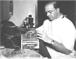
In the early 1940s, Dr. Frederic Mohs, Professor of Surgery at the University of Wisconsin, developed a form of treatment for skin cancer, which he called chemosurgery. (The word “chemosurgery” is derived from the words “chemical” and “surgery.”) The technique has undergone many refinements and came to be known as “Mohs Surgery” in honor of Dr. Mohs. Chemicals are no longer used. The term “Mohs Micrographic Surgery” was first published by Dr. Hanke in 1985 to indicate the tissue mapping (“graphic”) and specialized microscopic examination (“micro”) that are done. The shortened term Mohs Surgery is often used.
Dr. Hanke is a leader and innovator in the specialty of Mohs Surgery. He founded and directed the Mohs Surgery training programs at Indiana University and St. Vincent Hospital, Indianapolis. He currently directs the ACGME-accredited fellowship training program in Micrographic Surgery and Dermatologic Oncology at St. Vincent Hospital. Dr. Hanke serves on the ACGME Residency Review Committee for Dermatology which establishes standards for all training programs in the U.S. He was elected President of the American College of Mohs Surgery which has more than 1,000 members.
Dr. Hanke has contributed to the development of Mohs Surgery through his publications on technology for high-quality frozen section preparation and super wide field microscopy. The size and quality of his frozen sections are unprecedented thereby maximizing his ability to provide high cure rates for skin cancer.
What to Expect During Your Mohs Micrographic Surgery
Mohs Surgery Procedure
The surgery is performed as follows:
- The suspicious lesion is treated with a local anesthetic to eradicate feeling of discomfort in the area.
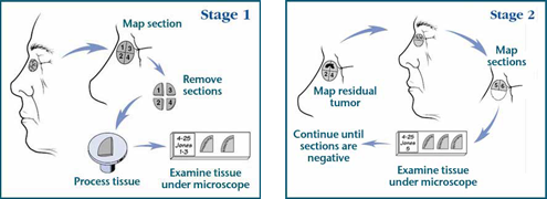
- The tumor is scraped using a sharp instrument called a curette to remove most of the visible cancer.
- A thin layer of tissue is then removed surgically and is carefully divided into pieces that will fit on a microscope slide.
Examination of Tissue
The tissue edges are marked with specially colored dyes; a careful diagram of the tissue removed is made and the tissue is frozen by the histo-technician. Thin slices are then made from the frozen tissue and examined by your surgeon under the microscope. Most bleeding from the procedure is controlled using pressure and other routine measures; occasionally a small blood vessel is encountered which must be tied using suture material or cauterized. A pressure dressing is applied, and the patient is asked to wait in the reception area while the slides are being processed.
Your Mohs surgeon will then examine the slides under the microscope to determine if cancer is still present. If cancer cells remain, he is able to judge the number of cells and the exact location. Another layer of tissue is then removed, and the procedure is repeated until the surgeon is satisfied that the entire base and side of the wound are cancer free. As well as ensuring total removal of the cancerous tissue, this process preserves as much normal, healthy skin as possible. The goal is to remove the cancerous tissue while minimizing loss of normal tissue.
Duration of Mohs Surgery
The removal of each layer of tissue takes one to two hours. Only 20 to 30 minutes of that time is spent in the actual surgical procedure. The remaining time is required for slide preparation and interpretation. Removal of two or three layers of tissue (called ‘stages’) is usually required to complete the surgery. Therefore, if begun early in the morning, it is generally completed in one day or part of one day. Occasionally, however, a tumor may be extensive enough to necessitate continuing surgery the following day.
Post-Operative Care for Mohs Surgery
Rate of Cure Following Mohs Procedures
Using Mohs surgery, the rate of success is very high, often 95 to 99 percent, even if other forms of treatment have failed. However, no one can guarantee a 100 percent cure rate.
Because normal tissue is preserved, Dr. Hanke is able to offer you the best possibility for a good cosmetic result. Every effort will be made to minimize the scar.
Schedule Your Skin Cancer Evaluation and Treatment at the Laser and Skin Surgery Center of Indiana
At the Laser and Skin Surgery Center of Indiana, we are experts in skin cancer. In addition to Mohs Micrographic Surgery, we also provide other skin cancer treatment options in our Indiana dermatologic surgery practice. Schedule a consultation today with Dr. Hanke to find out more about our various options and which skin cancer treatment is right for you.


